|
|
|
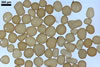
SPORES single in the soil; develop laterally on the neck of a sporiferous saccule; orange white (5A2) when young to reddish orange (7A8) when mature; globose to subglobose; (80-)183(-340) µm diam; sometimes irregular; 140-210 x 200-260 µm.
SUBCELLULAR STRUCTURE OF SPORES consists of a spore wall and two inner germination walls.
 |
 |
 |
 |
 |
 |
 |
In PVLG |
||||||
 |
 |
 |
|
|
In PVLG |
In PVLG+Melzer's reagent
|
|||
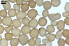
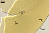
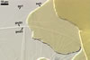
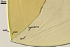
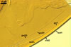
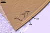
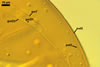
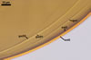
Layer 1, forming the spore surface, evanescent, hyaline, ca. 0.5 µm thick before disintegration, closely attached to layer 2, continuous with the wall of a sporiferous saccule, usually completely sloughed in mature spores.
Layer 2 laminate, smooth, orange white (5A2) to reddish orange (7A8), (1.2-)2.3(-3.2) µm thick.
Layer 3 laminate, hyaline, (0.7-)1.2(-1.7) µm thick, usually tightly adherent to layer 2, staining reddish orange (7B8) in Melzer's reagent.
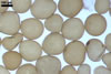
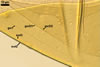
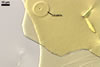
Germination wall 1 composed of a flexible to semiflexible, hyaline, (0.5-)0.8(-1.0) µm thick layer (gw1).
Germination wall 2 consists of two layers (gw2l1 and 2).
Layer 1 flexible, hyaline, covered with small granules, (0.7-)1.1(-1.2) µm thick.
Layer 2 plastic, 2-15 µm thick in PVLG, (0.5-)0.8(-1.0) µm thick and pastel pink (11A4) in Melzer’s reagent.
GERMINATION ORB. Not found.
 |
 |
In PVLG |
|
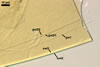
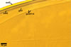
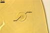
CICATRIX. Ca. 0.5-1.0 µm high, surrounds a pore, 17.5-35.0 µm diam.
MYCORRHIZAE. Acaulospora koskei has been associated in the field with vesicular-arbuscular mycorrhizae of Agrostis stolonifera L., Ammophila arenaria (L.) Link, and Juncus articulatus L. em. K. Richt. (Blaszkowski 1995).
DISTRIBUTION. In Poland, Acaulospora koskei has been found associated only with dune plants colonizing humid active deflation hollows of the Slowinski National Park (54º45’N, 17º26’E; Blaszkowski 1995; Tadych and Blaszkowski 2000).
NOTES. Under a dissecting microscope, spores of A. koskei most resemble in colour and size those of A. laevis and young, pale-coloured spores of A. capsicula. However, the spore wall layer 3 of only the former species stains in Melzer's reagent (Blaszkowski 1990, 1995; Morton 2002). Additionally, the germination wall 1 of both spores of A. capsicula and A. laevis consists of two layers, whereas that of A. koskei spores is 1-layered. Finally, the unique property of A. capsicula is the capsicum red colour of its mature spores.
REFERENCES
Blaszkowski J. 1990. Polish Endogonaceae. VII. Acaulospora capsicula sp. nov. Mycologia 82, 794-798.
Blaszkowski J. 1995. Acaulospora koskei, a new species in Glomales from Poland. Mycol. Res. 99, 237-240.
Morton J. B. 2002. International Culture Collection of Arbuscular and Vesicular-Arbuscular Mycorrhizal Fungi. West Virginia University. http://www.invam.caf.wvu.edu/.
Tadych M., Blaszkowski J. 2000. Arbuscular fungi and mycorrhizae (Glomales) of the Slowinski National Park, Poland. Mycotaxon 74, 463-483.