
In PVLG
Sieverd. & S. Toro
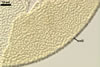
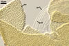
SPORES single in the soil; develop laterally on the neck of a sporiferous saccule; dull yellow (3B3) to Nankeen yellow (3B7); globose to subglobose; (90-)140(-165) µm diam.
SUBCELLULAR STRUCTURE OF SPORES consists of a spore wall and two inner germination walls.
|
|
|
|
|
|
|
In PVLG |
||||||
 |
In PVLG |
Layer 1 evanescent, hyaline, (0.8-)1.0(-1.2) µm thick, continuous with the wall of a sporiferous saccule, rarely present in mature spores.
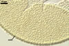
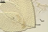
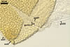
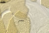
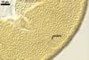
Layer 2 laminate, dull yellow (3B3) to Nankeen yellow (3B7), (4.5-)10.0(-12.0) µm thick, ornamented with labyrinthiform folds, 1.0-3.5 µm wide and 1.0-4.5 µm high when seen in a plane view and a cross view, respectively.
Layer 3 semiflexible, hyaline, (0.5-)0.7(-0.8) µm thick, rarely separable from layer 2.
Germination wall 1 consists of two tightly adherent hyaline, semiflexible layers (gw1l1 and 2), each 0.5-0.8 µm thick; these layers are tightly adherent to each other and, hence, difficult to see.
Germination wall 2 composed of two adherent layers (gw2l1 and 2).
Layer 1 flexible, hyaline, (0.5-)0.7(-1.0) µm thick, covered with small, <0.5 µm diam, granules scattering in crushed spores.
Layer 2 flexible, hyaline, (0.6-)1.1(-1.4) µm thick in PVLG; its staining reaction in Melzer's reagent was not examined.
GERMINATION ORB. Not observed.
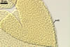
SPORIFEROUS SACCULE hyaline; globose to subglobose; 90-150 µm diam, with 1-layered wall, 0.8-1.7 µm thick; neck 50-80 µm long, tapering from 15.0-22.0 µm wide at the saccule to 12.0-19.0 µm wide at the spore attachment. Saccule usually collapses and is detached in mature spores.
 |
 |
In PVLG |
|
CICATRIX. A slightly raised, circular scar, 8.0-11.5 µm diam.
MYCORRHIZAE. In Poland, Ac. rehmii has been associated with vesicular-arbuscular mycorrhizal roots of Plantago lanceolata L. Many attempts to initiate sporulation of this fungus in both trap and one-species cultures failed. According to Sieverding and Toro (1987), Ac. rehmii produced vesicular-arbuscular mycorrhizae.
DISTRIBUTION. In Poland, spores of Ac. rehmii have been found only once. They have been recovered from among roots of P. lanceolata growing in a salt pan near Wladyslawowo (54º47’N, 18º26’E) at the beginning of the Hel Peninsula (Blaszkowski, unpubl.).
Acaulospora rehmii has originally been isolated from a crop field at Caicedonia, Valle de Cauca, Colombia (Sieverding and Toro 1987). This fungus has been associated with cassava, beans, sorghum, and Crotalaria species. Additionally, Sieverding and Toro (1987) found spores of Ac. rehmii in a culture containing a soil sample coming from the Centro de Pesquisa Agropecuária dos Cerrados, Brasilia D. F., Brazil. Recently, Moreira-Souza et al. (2003) confirmed the presence of this fungus in Brazil; Ac. rehmii spores were encountered among roots of Araucaria angustifolia (Bert.) O. Ktze growing in a reforested area in the State Park of Campos do Jordão, San Paulo. Wu and Liu (1995) isolated spores of Ac. rehmii from among roots of Phyllostachys pubescens Mazel growing in a garden in Chi-tou Experimental Station, National Taiwan University.
NOTES. Acaulospora rehmii is a species easy to recognize because of the unique labyrinthiform ornamentation of the wall layer 2 of its spores.
REFERENCES
Moreira-Souza M., Truem S. F. B., Gomes-da-Costa S. M. Cardoso E. J. B. N. 2003. Arbuscular mycorrhizal fungi associated with Araucaria angustifolia (Bert.) O. Ktze. Mycorrhiza 13, 211-215.
Sieverding E., Toro S. T. 1987. Acaulospora denticulata sp. nov. and Acaulospora rehmii sp. nov. (Endogonaceae) with ornamented spore walls. Angew. Bot. 61, 217-223.
Wu C.-G., Liu Y.-S., Hwuang Y.-L., Wang Y.-P., Chao C.-C. 1995. Glomales of Taiwan: V. Glomus chimonobambusae and Entrophospora kentinensis, spp. nov. Mycotaxon 53, 283-294.