DEVELOPMENT OF SPORES AND CHARACTERS OF MYCORRHIZAE
OF THE GENUS ACAULOSPORA
 |
 |
 |
 |
 |
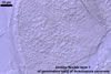
In PVLG
|
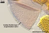
In water |
In PVLG |
In PVLG+Melzer's |
|
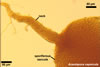
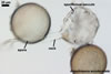
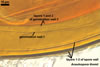
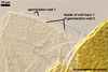
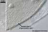
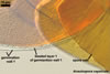
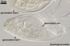
Once the spore wall is completely differentiated and spores have cased expansion, one or two discrete and separate flexible to semiflexible, hyaline inner germination walls are formed: the first one is one- or bilayered and the second one bi- or three-layered. In most species, the outer layer of the innermost germination wall is ornamented with beads (granular excrescences). In a few species, it is either ornamented with knobs or is smooth. The second or third layer of this wall usually stains intensively in Melzer's reagent; it rarely stains faintly or is nonreactive in this reagent.
 |
 |
 |
 |
 |
 |
In PVLG+Melzer's |
In PVLG
|
In PVLG+Melzer's |
In PVLG |
In PVLG+Melzer's |
|
 |
 |
 |
 |
In PVLG
|
In PVLG+Melzer's |
||
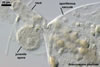
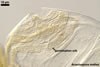
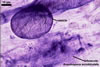
After both inner germination walls are fully differentiated, a “germination orb” is synthesized between the two germination walls. Spores of the genus Acaulospora germinate by germ tubes emerging from a plate-like germination orb formed by centrifugally rolled hypha (Blaszkowski 1994). The germ tubes penetrate through the spore wall.
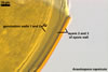
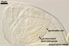
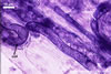
The mycorrhizae of Acaulospora spp. consist of (1) arbuscules with cylindrical or slightly flared trunks (similar to those of Glomus spp.), (2) irregular and knobby vesicles, and (3) straight and coiled intraradical hyphae with coils mostly concentrated at entry points (Morton 2000). All the structures usually have a patchy distribution and stain faint in trypan blue or other stains.
 |
 |
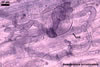 |
In roots of Plantago lanceolata L.
|
||
REFERENCES
Blaszkowski J. 1994. Polish Glomales 10. Acaulospora dilatata and Scutellospora dipurpurascens. Mycorrhiza 4, 173-182.
Morton J. B. 2000. International Culture Collection of Arbuscular and Vesicular-Arbuscular Mycorrhizal Fungi. West Virginia University.
Morton J. B., Benny G. L. 1990. Revised classification of arbuscular mycorrhizal fungi (Zygomycetes): a new order, Glomales, two new suborders, Glomineae and Gigasporineae, and two new families, Acaulosporaceae and Gigasporaceae, with an emendation of Glomaceae. Mycotaxon 37, 471-491.