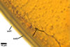
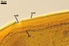
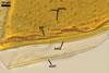
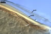
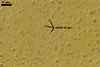
GERMINATION.
A germ tube develops from the germinal wall and penetrates the spore wall.
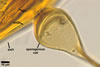
AUXILIARY
CELLS. No found.
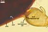
MYCORRHIZAE.
Not established yet. According to Bentivenga and Morton (1995),
the mycorrhizae of Gi. gigantea consisted of arbuscules and intraradical
hyphae staining darkly in trypan blue.
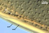
DISTRIBUTION.
In Poland, Gi. gigantea has been recorded under
different plants of dune sites of the Baltic Sea coast (Blaszkowski 1990,
1993, 1994; Tadych and Blaszkowski 2000).
This
species has commonly occurred in Ammophila breviligulata Fern.-dominated
dunes of the Atlantic coast of North America from Quebec to Virginia (Bergen
and Koske 1984; Dalpé 1989; Gemma and Koske 1989; Gemma et al. 1989;
Koske 1987; Koske and Halvorson 1981). Rose (1980) and Sylvia and Will (1988)
found this fungus in dunes of Florida. Gigaspora gigantea has also
inhabited dunes of Wisconsin (Koske and Tews 1987) and New South Wales, Australia
(Koske 1975).
NOTES.
Gigaspora gigantea is the type species of the genus Gigaspora
(Gerdemann and Trappe 1974). The most distinctive characters
of Gi. gigantea are the colour and size of its spores. The greenish
yellow spore colour is unknown elsewhere in the phylum Glomeromycota. Moreover,
this colour is associated with the spore contents rather than with the laminae
of the spore wall as in the other Gigaspora species. Of the five accepted
species of the genus Gigaspora (Bentivenga and Morton 1995), Gi.
gigantea produces largest spores. Nicolson and Gerdemann (1968) described
mature spores of this fungus as being as large as 812 µm diam.
Gigaspora gigantea
is one of only two species the genus Gigaspora found by the author of
this website in rhizosphere soils coming from different regions of Europe,
Africa, and Asia.
REFERENCES
Bentivenga S. P., Morton
J. B. 1995. A monograph of the genus Gigaspora, incorporating developmental
patterns of morphological characters. Mycologia 87, 719-731.
Bergen M., Koske R. E.
1984. Vesicular-arbuscular mycorrhizal fungi from sand dunes of Cape Cod,
Massachusetts. Trans. Br. Mycol. Soc. 83, 157-158.
Blaszkowski J. 1990.
Polish Endogonaceae IV. Gigaspora gigantea, Glomus deserticola,
and Glomus globiferum. Acta Mycol 26, 3-16.
Blaszkowski J. 1993.
Comparative studies of the occurrence of arbuscular fungi and mycorrhizae
(Glomales) in cultivated and uncultivated soils of Poland. Acta Mycol. 28,
93-140.
Blaszkowski J. 1994.
Arbuscular fungi and mycorrhizae (Glomales) of the Hel Peninsula, Poland.
Mycorrhiza 5, 71-88.
Dalpé Y. 1989.
Inventaire et repartition de la flore endomycorhizienne de dunes et de rivages
maritimes du Québec, du Nouveau-Brunswick et de la Nouvelle-Ecosse.
Naturaliste Can. (Rev. Ecol. Syst.) 116, 219-236.
Gemma J. N., Koske R.
E. 1989. Field inoculation of American beachgrass (Ammophila breviligulata)
with V-A mycorrhizal fungi. J. Environm. Manag. 29, 173-182.
Gemma J. N., Koske R.
E., Carreiro M. 1989. Seasonal dynamics of selected species of VA mycorrhizal
fungi in a sand dune. Mycol. Res. 92, 317-321.
Gerdemann J. W., Trappe
J. M. 1974. The Endogonaceae in the Pacific Northwest. Myc. Memoir 5, 1-76.
Koske R. E. 1975. Endogone
spores in Australian sand dunes. Can J Bot 53, 668-672.
Koske R. E. 1987. Distribution
of VA mycorrhizal fungi along a latitudinal temperature gradient. Mycologia
79, 55-68.
Koske R. E., Halvorson
W. L. 1981. Ecological studies of vesicular-arbuscular mycorrhizae in a barrier
sand dune. Can. J. Bot. 59, 1413-1422.
Koske R. E., Tews L.
L. 1987. Vesicular-arbuscular mycorrhizal fungi of Wisconsin sandy soils.
Mycologia 79, 901-905.
Nicolson T. H., Gerdemann
J. W. 1968. Mycorrhizal Endogone species. Mycologia 60, 313-325.
Rose S. L. 1980. Mycorrhizal
associations of some actinomycete nodulated nitrogen-fixing plants. Can. J.
Bot. 58, 1449-1454.
Sylvia D. M., Will M.
E. 1988. Establishment of vesicular-arbuscular mycorrhizal fungi and other
microorganisms on a beach replenishment site in Florida. Appl. Environm. Microbiol.
54, 348-352.
Tadych M., Blaszkowski
J. 2000. Arbuscular fungi and mycorrhizae (Glomales) of the Slowinski National
Park, Poland. Mycotaxon 74, 463-483.