Pacispora boliviana

Sieverd. & Oehl


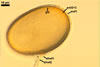
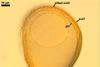
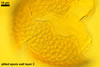
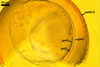
SPORES borne singly in the soil; blastically at the tip of mycorrhizal extraradical hyphae. Spores pale yellow (4A3) to sunflower yellow (4A5); globose to subglobose; 70-95 µm diam; or ellipsoid to irregular; 71-105 x (57-)62-80 µm; with a single subtending hypha.


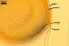
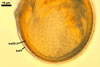
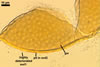
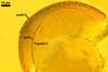
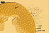
SUBCELLULAR STRUCTURE OF SPORES consists of a spore wall and an inner germination wall.
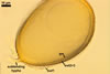
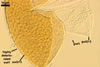
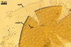
In PVLG
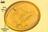
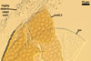
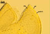
In PVLG+Melzer's reagent

Spore wall composed of three adherent layers (swl1-3).

Layer 1, forming the spore surface, sometimes laminate, hyaline, 1.3-2.3(-3.0) µm thick, of a smooth upper surface and a lower surface ornamented with processes projecting inward; the processes fill pits of the outer surface of layer 2; layer 1 gradually sloughs with age.

Layer 2 laminate, pale yellow (4A3) to sunflower yellow (4A5), 2.5-3.5(-4.5) µm thick, of the outer surface densely ornamented with shallow, usually pentagonal pits, 3.5-5.5 µm wide and 0.5-1.1 µm deep with dams 0.5-1.6 µm wide between the pits.

Layer 3 semi-flexible, hyaline to light yellow (4A4), ca. 0.5 µm thick, always tightly adherent to layer 2 and, hence, difficult to see.

Germination wall includes three hyaline layers (gwl1-3).

Layer 1 flexible, ca. 0.5 µm thick, easily separating from layer 2 in crushed spores.

Layer 2 coriaceous, 1.2-2.0 µm thick.

Layer 3 flexible, <0.5 µm thick, always tightly to layer 2, usually slightly wrinkled in spores vigorously crushed in PVLG.

In Melzer's reagent, none of the layers of both the spore wall and the inner germination wall stains.


GERMINATION SHIELD. Not found to date.


SUBTENDING HYPHA hyaline to yellowish white (4A2); straight or curved; cylindrical, occasionally slightly constricted at the spore base; 5-10 µm wide at the spore base.

In PVLG

Wall of subtending hypha hypha hyaline to yellowish white (4A2); 3.8-5.8(-7.5) µm thick at the spore base and frequently up to (5.0-)10-30(-60.0) µm thick below the spore base, then narrows to <0.5 µm thick; composed of two layers continuous with spore wall layers 1 and 2.

Pore usually closed at the spore base by a bridging septum formed by the innermost lamina of spore wall layer 2 and/or by the adherent spore wall layer 3.


GERMINATION. Not observed.


MYCORRHIZAE. Spores of P. boliviana have been associated with roots of plants of a trap culture with soil derived from a natural forest in the semi-arid, tropical Gran Chaco, District Santa Cruz, Bolivia (Oehl and Sieverding 2004). However, the characters of mycorrhizae of this fungus from one-species cultures have not been recognized to date.


DISTRIBUTION. The type of P. boliviana has been designated from spores of a trap culture with soil of the site presented above. It is the only site of the occurrence of this fungus so far reported in the literature.

NOTES. The morphological and biochemical properties of spores of P. boliviana presented above were prepared based on the original description of this fungus (Oehl and Sieverding 2004) and observations of its spores loaned from Dr. F. Oehl, Institute of Botany, University of Basel, Switzerland.

The only species of arbuscular fungi forming glomoid spores of the outer surface of the structural laminate layer of their wall ornamented with pits is Gl. multiforum Tadych & Blaszk. Compared with spores of P. boliviana, spores of the latter fungus are much darker [deep yellow (4A8) to brown (6E8) vs. pale yellow (4A3) to sunflower yellow (4A5) in P. boliviana] and larger [(170-)215(-230) µm diam vs. 70-95 µm diam; Blaszkowski 2003; Blaszkowski and Tadych 1997; Oehl and Sieverding 2004]. However, the most important difference between these fungi is the lack of the inner germination wall of spores of P. boliviana in spores of Gl. multiforum.

Of the other species of the Pacispora, none has spores with an ornamented structural laminate layer. In Pacispora spp. producing ornamented spores, the ornamentation occurs only on the surface of the outermost spore wall layer and consists of warts or ridges directed outwards the spore, as, e. g., in P. scintillans (Blaszkowski 2003; Oehl and Sieverding 2004).


REFERENCES

Blaszkowski J. 2003. Arbuscular mycorrhizal fungi (Glomeromycota), Endogone, and Complexipes species deposited in the Department of Plant Pathology, University of Agriculture in Szczecin, Poland. http://www.agro.ar.szczecin.pl/~jblaszkowski/.

Blaszkowski J., Tadych M. 1997. Glomus multiforum and G. verruculosum, two new species from Poland. Mycologia 89, 804-811.

Oehl F., Sieverding E. 2004. Pacispora, a new vesicular arbuscular mycorrhizal fungal genus in the Glomeromycetes. J. Appl. Bot. 78, 72-82.