 |
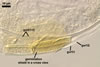 |
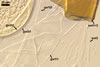
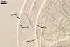
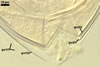
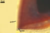
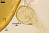
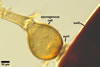
In PVLG |
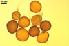
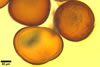
GERMINATION
SHIELD cardioid to irregular; pale yellow (3A3); 45.0-67.5 x
80.0-94.0 µm; with deep folds
partitioning 5-11 lobes with nicked margins formed by shallow incisions; formed
on germinal wall 2. One to three, hyaline to pale yellow (3A3), 1.8-5.4 µm
diam germ tubes emerge from the germination shield.
 |
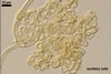 |
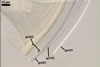
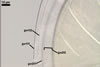
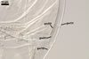
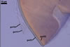
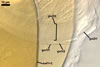
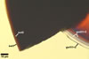
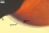
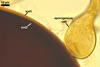
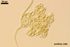
In PVLG |
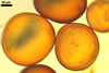
AUXILIARY
CELLS rarely single in the soil, usually in loose clusters of
2-12; hyaline to pale yellow (3A3); lobed to irregular; 15.0-32.5 x 22.5-32.5
µm; with knobby projections, 2.5-11.0 x 3.0-10.0 µm; produced on
coiled hyphae, 3.0-5.5 µm diam, concolorous with auxiliary cells.
MYCORRHIZAE.
Scutellospora armeniaca has originally been described
from spores recovered from among vesicular-arbuscular mycorrhizal roots of
Artemisia campestris L. colonizing maritime dunes adjacent to Chalupy
(54o46’N, 18o31’E) located on the Hel Peninsula in northern Poland
(Blaszkowski 1992). Later, this fungus has been found associated with vesicular-arbuscular
mycorrhizal roots of many other dune and non-dune plant species (Blaszkowski
1992, 1993; Blaszkowski et al. 2002a, b; Tadych and Blaszkowski 2000a, b).
DISTRIBUTION.
In
Poland, S. armeniaca has occurred in dune sands of the Vistula Bar
(54º21’N, 19º14’E; Blaszkowski et al. 2002a), the Gdansk coast
(Blaszkowski 1992, 1993), and the Slowinski National Park (54º45’N,
17º26’E; Tadych and Blaszkowski 2000a). This species has been the most
frequently occurring arbuscular fungus in soils of the Bledowska Desert (50º22’N,
19º34’E; Blaszkowski et al. 2002b). Additionally, spores of S. armeniaca have been revealed in soils of the Tuchola Forests (53º46’N, 17º42’E-53º40’N,
17º54’E; Tadych and Blaszkowski 2000b).
No report
exists of the finding of S. armeniaca in other regions of the world.
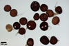
NOTES. Spores of S. armeniaca are most likely to be confused with those of S. arenicola, S. castanea, S. erythropa, and S. hawaiensis. All these species produce spores similar in colour (Koske and Gemma 1995; Koske and Halvorson 1989; Koske and Walker 1984; Morton 2002; Stürmer and Morton 1999; Walker et al. 1993). However, even the smallest spores of S. castanea [(285-)298(310) µm diam when globose; Walker et al. 1993] are larger than the largest spores of S. armeniaca [(140-)196(-240) µm diam; Blaszkowski 1992]. Spores of S. arenicola [(160-)230(-360) x (120-)220(-310) µm; Koske and Halvorson 1989], S. erythropa [(160-)240(-320) µm diam; Morton 2002], and S. havaiensis [(200-)240(-360) x (180-)230(-290) µm; Koske and Gemma 1995] may also be much larger than those of the fungus discussed here.
The main differences between these fungi inhere in the subcellular structure of their spores, especially in the number and the composition of the inner germinal walls. Of the species compared here, a third flexible spore wall layer was revealed only in S. armeniaca. A similar layer was found in the spore wall of, e. g., S. heterogama and S. rubra (Stürmer and Morton 1999). This layer usually is very thin and tightly adheres to a structural laminate spore wall layer, and hence is difficult to see, especially in field-collected spores.
Scutellospora castanea and S. erythropa diverge from the other species considered here in the number of germinal walls. In S. castanea, only one 2-layered germinal wall is present (Walker et al. 1993), and S. erythropa differentiates three germinal walls (Morton 2002; vs. two, 2-layered germinal walls in the other species).
REFERENCES
Blaszkowski J. 1992.
Scutellospora armeniaca, a new species in Glomales (Zygomycetes)
from Poland. Mycologia 84, 939-944.
Blaszkowski J. 1993.
The occurrence of arbuscular fungi and mycorrhizae (Glomales) in plant communities
of maritime dunes and shores of Poland. Bull. Pol. Ac. Sci. Biol. Sci. 41,
377-392.
Blaszkowski J., Adamska
I., Czerniawska B. 2002a. Arbuscular mycorrhizal fungi (Glomeromycota) of
the Vistula Bar. Acta Mycol. 37, 39-62.
Blaszkowski J., Tadych
M., Madej T. 2002b. Arbuscular mycorrhizal fungi (Glomales, Zygomycota) of
the Bledowska Desert, Poland. Acta Soc. Bot. Pol. 71, 71-85.
Koske R. E., Gemma J.
N. 1995. Scutellospora hawaiiensis: a new species of arbuscular mycorrhizal
fungus from Hawaii. Mycologia 87, 678-683.
Koske R. E., Halvorson
W. L. 1989. Scutellospora arenicola and Glomus trimurales:
two new species in the Endogonaceae. Mycologia 81, 927-933.
Koske R. E., Walker
C. 1984. Gigaspora erythropa, a new species forming arbuscular mycorrhizae.
Mycologia 76, 250-255.
Morton J. B. 2002. International
Culture Collection of Arbuscular and Vesicular-Arbuscular Mycorrhizal Fungi.
West Virginia University. http://www.invam.caf.wvu.edu/.
Stürmer S. L.,
Morton J. B. 1999. Scutellospora rubra, a new arbuscular mycorrhizal
species from Brazil. Mycol. Res. 103, 949-954.
Tadych M., Blaszkowski
J. 2000a. Arbuscular fungi and mycorrhizae (Glomales) of the Slowinski National
Park, Poland. Mycotaxon 74, 463-483.
Tadych M., Blaszkowski
J. 2000b. Arbuscular mycorrhizal fungi of the Brda river valley in the Tuchola
Forests. Acta Mycol. 35, 3-23.
Walker C., Gianinazzi-Pearson
V., Marion-Espinasse H. 1993. Scutellospora castanea, a newly described
arbuscular mycorrhizal fungus. Cryptog. Mycol. 14, 279-286.