Scutellospora fulgida

Koske & C. Walker


SPORES formed singly in the soil; origin blastically at the tip of a bulbous sporogenous cell; yellowish white (4A2) to cream (4A3); globose to subglobose; (165-)229(-280) µm diam.


SUBCELLULAR STRUCTURE OF SPORES consists of a spore wall and one inner germinal wall.

In PVLG
In PVLG+Melzer's reagent

Spore wall composed of two layers (swl1-2).

Layer 1, forming the spore surface, permanent, smooth, pale yellow (3A3) to light orange (5A5), (1.2-)2.0(-3.2) µm thick, usually tightly adherent to layer 2, sometimes slightly separated from it in vigorously crushed spores.

Layer 2 laminate, smooth, yellowish white (4A2) to cream (4A3), (7.6-)9.7(-13.0) µm thick.

Germinal wall comprises two flexible, smooth layers (gwl1 and 2).

Layer 1 ca. 0.5 µm thick, adherent to layer 2, but frequently wrinkled in crushed spores and, thereby, giving the inner wall a rugose or blistered appearance and making this layer more visible.

Layer 2 0.8-1.5 µm thick.

In Melzer's reagent, the spore wall layers 1 and 2 stain orange white (5A2) to copper (7C8) and pale yellow (3A3) to reddish orange (7B7), respectively, and both germinal wall layers remain nonreactive.


SPOROGENOUS CELL formed terminally on a sparsely septate hypha continuous with an extraradical mycorrhizal hypha; ovoid to clavate; (38.7-)44.6(-53.2) µm wide; cream (4A3) to light orange (5A5), usually with a hyphal peg.
In PVLG
In PVLG+Melzer's reagent
Wall of sporogenous cell composed of two permanent layers (layers 1 and 2) continuous with spore wall layers 1 and 2.

Layer 1 pale yellow (3A3) to light orange (5A5), (1.7-)2.4(-3.2) µm thick.

Layer 2 cream (4A3) to light yellow (4A4), (1.5-)2.6(-3.9) µm thick at the spore base. Both layers always tightly adherent to one another and frequently difficult to observe.


GERMINATION SHIELD cardioid; pale yellow (3A3); 60-120 x 110-190 µm, of a more or less incised border; ornamented with widely, usually unequally dispersed warts, 1.5-2.5 x 0.5-1.5 µm; positioned on the upper surface of the inner germinal wall. One to three germ tubes or germ tube initials emerge from the germination shield. In the soil, germ hyphae are straight or branched.

In PVLG
In PVLG+Melzer's reagent

In PVLG
In PVLG+Melzer's

AUXILIARY CELLS borne in the soil, in clusters of 6-10; hyaline to cream (4A3); globose to irregular; 18-28 x 23-39 µm; with knobby projections; produced on straight or coiled hyphae; 2.5-8.0 µm diam; concolorous with auxiliary cells.


MYCORRHIZAE. The presence of mycorrhizae in field-collected root samples was not determined. Many attempts to establish one-species cultures of S. fulgida using its spores extracted from trap cultures failed.


PHYLOGENETIC POSITION. According to De Souza et al. (2005), S. fulgida belongs in the clade C of the family Gigasporaceae and its closest relative is S. castanea.


DISTRIBUTION. The spores of S. fulgida showed here were extracted from six trap cultures with rhizosphere soils and root fragments collected under plants colonizing maritime dunes of Portugal (four samples), Italy and Oman (one sample each). The occurrence of arbuscular fungi in the field soil-root mixtures was not determined. The field samples from Italy came from under Ammophila arenaria (L.) Link and were collected on 11 October 2002. The exact sites and dates of collections of the soil and root samples, as well as the host plants sampled in Portugal and Oman are unknown. The trap cultures with these samples were established on 8 March 2004 and 30 June 2003, respectively, i. e., some days after they arrived to the laboratory of the author of this website. The arbuscular fungi accompanying S. fulgida in the cultures representing Italy were Acaulospora scrobiculata Trappe, Intraspora schenckii (Sieverd. & S. Toro) Oehl & Sieverd., Glomus aurantium Blaszk., V. Blanke, C. Renker & F. Buscot, Gl. constrictum Trappe, Gl. versiforme (P. Karsten) S.M. Berch, an undescribed Glomus 169, and S. persica (Koske & C. Walker) C. Walker & F.E. Sanders. The only arbuscular fungus co-occurring with S. fulgida in the Portugal culture was an undescribed Glomus 172, and the culture with the rhizosphere soil and root mixture of Oman still contained spores of Gl. fasciculatum (Thaxt.) Gerd. & Trappe emend. C. Walker & Koske and S. persica.

The type of S. fulgida comes from field-collected spores isolated from under A. breviligulata Fern. colonizing maritime dunes of the Seashore State Park in Virginia, U.S.A. (Koske and Walker 1986). This fungus has also been found in other soil samples taken from under A. breviligulata, Solidago sempervirens L., and Uniola paniculata L. growing in dunes extending from New Jersey to Virginia (Koske 1987). Sylvia and Will (1988) found spores of S. fulgida associated with U. paniculata and Panicum sp. growing in soils of a beach replenishment site in Florida. Additionally, S. fulgida has been reported to occur under Triticum aestivum L. cultivated in Argentina (Schalamuk et al. 2006) and in soils of China (Gai et al. 2006).


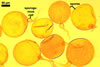
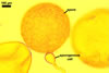
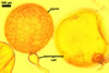
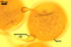
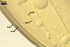
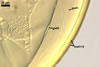
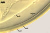
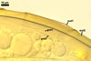
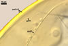
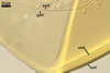
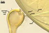
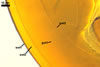
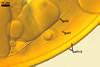
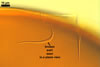
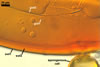
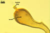
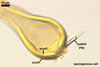
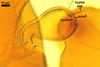
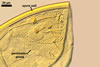
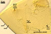
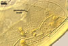
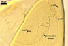
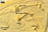
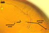
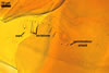
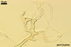
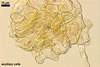
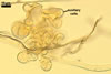
NOTES. The distinctive morphological characters of S. fulgida are its light-coloured and smooth spores having only one inner germinal wall. The last property keys this fungus into a monophyletic group still comprising S. castanea, S. coralloidea, S. gregaria, S. persica, and S. verrucosa.

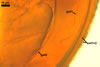
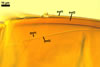
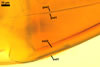
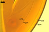
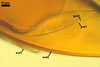
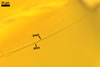
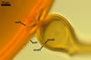
Four spore characters readily separate the species listed above. First, spores of S. fulgida are much lighter-coloured than those of the other species compared here (cream to light orange in S. fulgida vs. from pale straw to orange brown in S. verrucosa to red brown to dark brown in S. gregaria; Morton 1995, 2002). Second, in contrast to the smooth spores of S. castanea (Walker et al. 1993) and S. fulgida, the upper spore surface of the other species is ornamented with warts (Morton 1995, 2002). However, S. fulgida and S. castanea markedly differ in colour and size of spores. The darkest spores of the former fungus are of a yellow shade, and mature spores of the latter species are brown (Walker et al. 1993). Third, although the lower size range of globose spores of S. fulgida and S. castanea overlaps, the largest spores of S. fulgida (280 µm diam) are much smaller than the greatest spores of S. castanea (up to 372 µm diam; Walker et al. 1993). Spores of the other species discussed here may also attain a much higher size than those of S. fulgida (384 µm diam in S. persica to 480 µm diam in S. gregaria; Morton 1995). Fourth, similarly as in S. castanea, the warts ornamenting the germination shield of S. fulgida spores are much lower and less densely dispersed on its upper surface compared with those ornamenting the germination shield of the other species. This also causes the germination shields of the former two species to be relatively more flexible, as Morton (1995) concluded.


REFERENCES

De Souza F. A., Declerck S., Smit E., Kowalchuk G. A. 2005. Morphological, ontogenetic and molecular characterization of Scutellospora reticulata (Glomeromycota). Mycol. Res. 109, 697-706.

Gai J. P., Christie P., Feng G., Li X. L. 2006. Twenty years of research on biodiversity and distribution of arbuscular mycorrhizal fungi in China : a review. Mycorrhiza 16, 229-239.

Koske R. E. 1987. Distribution of VA mycorrhizal fungi along a latitudinal temperature gradient. Mycologia 79, 55-68.

Koske R. E., Walker C. 1986. Species of Scutellospora (Endogonaceae) with smooth-walled spores from maritime sand dunes: two new species and a redescription of the spores of Scutellospora pellucida and Scutellospora calospora. Mycotaxon 27, 219-235.

Morton J. M. 1995. Taxonomic and phylogenetic divergence among five Scutellospora species based on comparative developmental sequences. Mycologia 87, 127-137.

Morton J. B. 2002. International Culture Collection of (Vesicular) Arbuscular Mycorrhizal Fungi. West Virginia University: http://www.invam.caf.wvu.edu/.

Schalamuk S., Velazquez S., Chidichimo H., Cabello M. 2006. Fungal spore diversity of arbuscular mycorrhizal fungi associated with spring wheat: effect of tillage. Mycologia 98, 16-22.

Sylvia D. M., Will M. E. 1988. Establishment of vesicular-arbuscular mycorrhizal fungi and other microorganisms on a beach replenishment site in Florida. Appl. Environ. Microbiol. 54, 348-352.

Walker C., Gianinazzi-Pearson V., Marion-Espinasse H. 1993. Scutellospora castanea, a newly described arbuscular mycorrhizal fungus. Cryptog. Mycol. 14, 279-286.