(Trappe) R.T. Almeida & N.C. Schenck
 |
 |
 |
 |
 |
 |
In PVLG |
|||||
(Trappe) R.T. Almeida & N.C. Schenck
|
|
|
|
|
|
In PVLG |
|||||
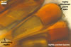
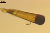
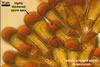
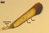
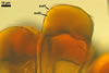
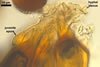
Sporocarps orange (5B8) to brown (6E8); globose to subglobose; 320-750 x 340-780 µm diam; minutely verrucose from exposed tips of spores; without a peridium and usually with one to four monohyphal stalks.
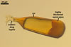
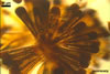
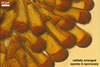
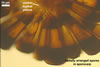
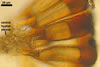
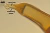
Spores orange (5B8) to brown (6E8); clavate to subcylindric, tapering towards a subtending hypha, (17.5-)41.0(-57.5) µm wide at the apex, (7.5-)10.0(-13.8) µm wide at the spore base, (27.5-)118.5(-150.0) µm long; formed radially in a single, tightly packed layer around a central plexus of hyphae.
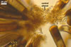
SUBCELLULAR STRUCTURE OF SPORES consists of one wall composed of two layers (swl1 and 2).
 |
 |
 |
 |
 |
 |
In PVLG |
In PVLG+Melzer's |
||||
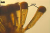
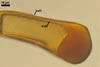
Layer 1, forming the spore surface, evanescent, hyaline, (0.5-)0.7(-0.9) µm thick, rarely present in mature spores.
Layer 2 laminate, orange (5B8) to brown (6E8), (2.2-)3.4(-4.7) µm thick on the sides, (22.5-)36.4(-50.0) µm thick at the apex.
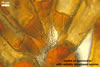
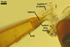
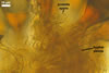
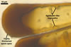
SUBTENDING HYPHA straight or recurved; cylindrical to slightly flared; (4.9-)12.0(-19.6) µm wide at the spore base.
 |
 |
 |
In PVLG |
In PVLG+Melzer's |
|
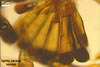
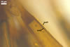
Wall of subtending hypha orange (5B8) to reddish orange (7B8); (4.7-)7.6(-8.6) µm thick at the spore base; composed of two layers continuous with the spore wall layers 1 and 2.Pore (1.2-)1.5(-1.7) µm diam, open or occluded by thickening of the inner laminate layer (layer 2) of the subtending hyphal wall, less frequently by a transverse septum connecting the inner surface of layer 2 of the subtending hyphal wall.
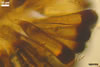
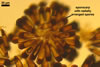
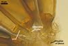
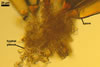
CENTRAL PLEXUS 110-450 µm diam, composed of tightly interwoven, yellowish white (4A2) to yellowish orange (4B7), septate, thin-walled hyphae, ca. 2.5-7.0 µm wide.
 |
 |
 |
 |
 |
In PVLG |
||||
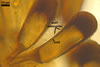
Neither spores nor the central plexus of hyphae stain in Melzer's reagent.
 |
In PVLG |
GERMINATION. A germ tube emerges from the lumen of the subtending hypha.
MYCORRHIZAE. Unknown.
PHYLOGENETIC POSITION. Unknown.
DISTRIBUTION. The type of Gl. clavisporum has been selected from sporocarps found among roots of grasses and forbs growing 40 km south of Catemaco near Hueyapan, Veracruz, Mexico (Trappe 1977). In Mexico, Gl. clavisporum has also been associated with roots of Zea mays L. (Estrada-Torres et al. 1992) and plants of a tropical lowland rain forest (Guadarrama and Alvarez-Sanchez 1999). Additionally, this fungus has been identified in cultivated and uncultivated sites of, e. g., Brazil (Almeida and Schenck 1990), Cameroon (Musoko et al. 1994), Israel (Dodd and Krikun 1984), Pakistan (Iqbal and Perveen 1980), China (Gai et al. 2006), and Taiwan (Wu 1993).
NOTES. Glomus clavisporum presented here was characterized based on results of studies of specimens provided by Dr F. Oehl, Institute of Botany, University of Basel, Switzerland, (slides no. ........) and descriptions of this fungus published by Almeida and Schenck (1990), Trappe (1977), and Wu (1993b).
Except for the characters of the spore wall and the lack of any hyphal stalk in the sporocarps provided, all the other properties of this fungus generally fitted those given by Almeida and Schenck (1990), Trappe (1977), and Wu (1993b).
As results from the descriptions of Almeida and Schenck (1990), Trappe (1977) and Wu (1993b), the spore wall of Gl. clavisporum consists of only one, coloured, laminate layer. However, in a part of the spores examined by me, the laminate layer was covered with a thin, colourless layer. In a few spores, it seemed to be relatively rigid and was intact. In other spores, this layer was more or less deteriorated or was absent at all. Thus, by definition (Walker 1983), it is of the type of sloughing layers.
Glomus clavisporum has originally been described as Sclerocystis clavispora Trappe (Trappe 1977). Considering differences in the mode of sporocarp formation and spore development, Almeida and Schenck (1990) remained in the genus Sclerocystis Berk. & Broome emend. R.T. Almeida & N.C. Schenck only S. coremioides Berk. & Broome. Of the 13 other described Sclerocystis spp., five were transferred to the genus Glomus Tul. & C. Tul. [as Gl. clavisporum, Gl. liquidambaris (C.G. Wu & Z.C. Chen) R.T. Almeida & N.C. Schenck, Gl. rubiforme (Gerd. & Trappe) R.T. Almeida & N.C. Schenck, Gl. sinuosum (Gerd. & B.K. Bakshi) R.T. Almeida & N.C. Schenck, Gl. taiwanense (C.G. Wu & Z.C. Chen) R.T. Almeida & N.C. Schenck], and eight were considered to be synonymous. Wu (1993b) rejected the changes of Almeida and Schenck (1990) and reverted to the previous classification of Gerdemann and Trappe (1974). However, phylogenetic analyses of the 18S ribosomal subunit of S. coremioides and S. sinuosa Gerd. & Bakshi have shown them to be members of a monophyletic clade comprising, e. g., Gl. intraradices N.C. Schenck & G.S. Sm. and Gl. mosseae (Nicol. & Gerd.) Gerd. & Trappe (Redecker et al. 2000). Consequently, S. coremioides was renamed Gl. coremioides (Berk. & Broome) D. Redecker & J.B. Morton and the genus Sclerocystis stopped to exist.
The distinctive characters of Gl. clavisporum are its relatively large sporocarps lacking a peridium and consisting of clavate, darkly coloured spores radially arranged, side by side, in a single, tightly packed layer around a central plexus of hyphae.
Of the known fungi of the Glomeromycota forming glomoid spores, Gl. clavisporum most resembles Gl. taiwanense. Both species form sporocarps lacking a peridium and their spores are similarly shaped and arranged in the sporocarp. Moreover, the spore wall of the two species consists of two layers of identical phenotypic and biochemical properties (Almeida and Schenck 1990; Blaszkowski, pers. observ.; Wu 1993b). However, both the sporocarps and spores of Gl. clavisporum are much larger [sporocarps: 320-750 x 340-780 µm; Almeida and Schenck 1990; spores: (80-)120-160(-185) x (20-)25-40(-50) µm or 27.5-150.0 x 7.5-57.5 µm; Almeida and Schenck 1990; Blaszkowski, pers. observ., respectively] than those of Gl. taiwanense [sporocarps: 200-300 x 180-280 µm; spores: 40-85(-105) x (17.5-)22-42.5(-55) µm; Almeida and Schenck 1990; Wu 1993b; Wu and Chen 1987].
 |
In PVLG |
In a few spores, so called lignituber-like ingrowths occurred earlier described from, e. g., other Glomus species, including members of the former genus Sclerocystis (Gerdemann and Bakshi 1977; Iqbal and Perveen 1980; Mosse and Bowen 1968). The ingrowths are characterized by having a narrow central channel and probably are the result of invasion of spores by soil microparasites (Koske 1985).
REFERENCES
Almeida R. T., Schenck N. C. 1990. A revision of the genus Sclerocystis (Glomaceae, Glomales). Mycologia 82, 703-714.
Dodd J. C., Krikun J. 1984. Observations on endogonaceous spores in the Negev Desert. Trans. Br. Mycol. Soc. 82, 536-540.
Estrada-Torres A., Varela L., Hernandez-Cuevas L., Cavito M. E. 1992. Algunos hongos micorrizicos arbusculares del estado de Tlaxcala, México. Rev. Mex. Mic. 8, 85-110.
Gai J. P., Christie P., Feng G., Li X. L. 2006. Twenty years of research on biodiversity and distribution of arbuscular mycorrhizal fungi in China: a review. Mycorrhiza 16, 229-239.
Gerdemann J. W. and Bakshi B. K. 1976. Endogonaceae of India : two new species. Trans. Brit. Mycol. Soc. 66, 340-343.
Gerdemann J. W., Trappe J. M. 1974. The Endogonaceae in the Pacific Northwest. Myc. Memoir 5, 1-76.
Guadarrama P., Alvarez-Sanchez F. J. 1999. Abundance of arbuscular mycorrhizal fungi spores in different environments in a tropical rain forest, Veracruz, Mexico. Mycorrhiza 8, 267-270.
Iqbal S. H., Perveen B. 1980. Some species of Sclerocystis (Endogonaceae) from Pakistan. Trans. Mycol. Soc. Japan 21, 57-63.
Koske R. E. 1985. Glomus aggregatum emended: A distinct taxon in the Glomus fasciculatum complex. Mycologia 77, 619-630.
Mosse B., Bowen G. D. 1968. A key to the recognition of some Endogone spore types. Trans. Br. Mycol. Soc. 51, 469-483.
Musoko M., Last F. T., Mason P. A. 1994. Populations of spores of vesicular-arbuscular mycorrhizal fungi in undisturbed soils of secondary semideciduous moist tropical forest in Cameroon. Forest Ecol. Manag. 63, 359-377.
Redecker D., Morton J. B., Bruns T. D. 2000. Molecular phylogeny of the arbuscular mycorrhizal fungi Glomus sinuosum and Sclerocystis coremioides. Mycologia 92, 282-285.
Trappe J. W. 1977. Three new Endogonaceae: Glomus constrictus, Sclerocystis clavispora, and Acaulospora scrobiculata. Mycotaxon 6, 359-366.
Walker C. 1983. Taxonomic concepts in the Endogonaceae: spore wall characteristics in species descriptions. Mycotaxon 18, 443-455.
Wu C.-G. 1993. Glomales of Taiwan: IV. A monograph of Sclerocystis (Glomaceae). Mycotaxon 49, 327-349.
Wu C.-G., Chen Z.-C. 1987. The Endogonaceae of Taiwan II: Two new species of Sclerocystis from Taiwan. Trans. Mycol. Soc. R.O.C. 2, 73-83.