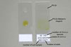
To make diagnostic slides with fungi freely occurring on the surface of different plant parts or growing in agar cultures, their sporophores with spores and mycelium were collected by means of the end of a needle and placed in a drop of water, lactic acid, polyvinyl alcohol/lactic acid/glycerol (PVLG; Koske and Tessier 1983), or a mixture of PVLG and Melzer’s reagent (1:1 v/v), and then covered with a cover slip. Occasionally, thin longitudinal and transverse cuttings were taken from the plant parts affected to show the fungal structures occurring both on their surface, e. g. sporophores, and inside them, e. g., haustoria.
 |
 |
 |
 |
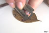
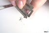
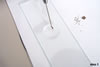
In case of fungi forming fruit bodies, e. g., apothecia or pycnidia, or other spore-bearing structures, e. g., uredinia, using a safety razor, thin cuttings were taken from transverse and/or longitudinal sections of the structures and the organ of its plant host. Four steps were usually followed to obtain such cuttings: (1) a relatively large (0.5-1 x ca. 0.5 cm) fragment was cut off from the place of the plant organ, e. g., a leaf, where the fungal structure intended to observe occurred; (2) as thin as possible cuttings were cut off from the vertical or horizontal plane of this fragment and the fungal structure present, e. g., from the vertical plane of the center of a pycnidium to show its ostiolum and wall with conidia-bearing cells; (3) the cut off cuttings were transferred by means of the wetted end of a needle to a drop of one the mounting media mentioned above, and (4) covered with a cover glass at a ca. 45o angle and observed.
In studies of arbuscular mycorrhizal fungi of the phylum Glomeromycota, intact and crushed spores mounted in PVLG and a mixture of PVLG and Melzer’s reagent (1:1 v/v) were used to make their both impermanent and permanent vouchers. Most spores of the genera Glomus and Gigaspora were crushed by applying light to moderate pressure on the cover slip with the end of a needle. Spores of the genera Acaulospora, Archaeospora, Entrophospora, Pacispora, and Scutellospora were frequently also first placed in a drop of lactic acid and then slightly crushed with the end of a needle. Such crushed spores were stored for 12-24 h and then used to prepare the slides, as described below. The earlier crushing loosens adherent layers, especially those of germination walls and, thereby, makes them to be better visible.