|
|
|
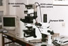 |
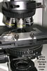
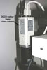
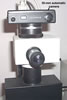
Microphotographs of of the fungal structures and mycorrhizae presented in this website were taken using a SONY 3CCD DXC-390 video camera. The camera may be positioned on either an OLYMPUS BX 50 light microscope equipped with differential interference contrast optics or an OLYMPUS SZX9 dissecting microscope. This system is connected with a Pentium III computer containing an Olympus Micro Image 128 capture KitTM version 3.1.1. Colour hardcopies of images are printed by means of the CP700DS MITSUBISHI dye-sublimation printer. Additionally, each of these microscopes has an adapter to accommodate a 35-mm automatic camera.
All macroscopic fungal structures, agar cultures, disease symptoms, etc. were photographed by using the Olympus digital camera Camedia 5050.
At present, about 10000 colour digital photographs of structures of saprotrophic and pathogenic fungi, as well as of spores and mycorrhizae of fungi of the phylum Glomeromycota coming from living cultures and field collections are stored on a hard disc of our computer. They are jpeg compressed. All of them are also copied to CD discs. Additionally, negatives of ca. 2000 of black and white and colour photographs are maintained. The most representative hard copies are stored in cardboard boxes.