|
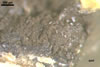
SORI developing from a locally condensed mycelium located in the outer parenchymal cells of a olive yellow (2D8) to yellowish brown (5F8), 8-14 x 16-23 mm, knobby outgrowths of a roughed surface, covered with a dense tomentum of the plant host, on stems, especially directly above nodes. Later, the hyphal mycelium converts into a gel that then develops into sori with spores.
 |
 |
 |
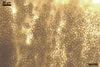
SPORES (s) subglobose, 6.1-9.6 µm diam, sometimes elliptic, 4.4-6.9 x 8.1-12.7 µm, of a greyish orange (5B3) to brownish yellow (5C7) colour.
Wall ornamented with warts of a different size.
PLANT HOST AND DISTRIBUTION. In Poland, U. trichophora has been found associated with Echinochloa crus-galli (L.) Beauv. growing in Rakowice Wielkie (51°08'N, 15°34'E; Madej et al. 2001).
According to Vánky (1994), this fungus has a worldwide distribution.
IN VITRO DEVELOPMENT. The germinability of spores on potato dextrose agar (PDA), Sabouraud dextrose agar (SDA) and in water after 6 months of storage in dry conditions ranged from 14 to 35%. The percent of germination was higher when the spores occurred in aggregates. Single spores germinated infrequently. On PDA, SDA, and in water, the spores produced a promycelium after 16-24 h. The promycelium usually was one-celled; sometimes it was branched at the spore base. Sporidia formed at the tip of the promycelium by budding. Setchall and Brefeld (in Vánky 1974) informed that the promycelium of U. trichophora is two-celled. The promycelium subsequently developed into colonies.
On PDA, after 3 weeks of growth, U. trichophora produced yellowish white (4A2) to orange white (5A2), compact, surface colonies of a diameter of 3-3.5 cm with a cream (4A3) to butter yellow (4A5) reverse. The aerial mycelium of these colonies formed branched, hyaline hyphae, 2-2.5 µm wide, consisting of longitudinally ellipsoidal, hyaline cells with a granular content. With age, the colonies became leathery, and their aerial mycelium produced numerous globose to oval conglomerations due to the fragmentation of the hyphae.
On SDA, the colonies of U. trichophora were compact, powdery to slimy, of an orange (5B8) to yellowish brown (5D8) colour; with age, their aerial mycelium also fragmented into globose, hyaline to flavus pieces.
The attempts to infect E. crusgalli by both the inoculation of seeds with spores and seedlings with a water suspension of sporidia failed.
NOTES. The properties of the spores presented here correspond with those given by Brandenburger (1985), Blumer (1963), and Vánky (1994).
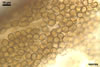
The sori were covered with 6-15 layers of parenchymal cells deformed due to hypertrophy and hyperplasia. The cells were almost hyaline, lacked cytoplasm, and usually contained remnants of the mycelium. The integuments, even wider than 50 µm, burst when spores were mature.
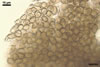
The living cells neighbouring the sori were colonized by hyphae developing only intracellularly. The hyphae were hyaline, 2.0-3.5 µm wide, and had many short branches ended with haustoria of different shapes. According to Vánky (1994), U. trichophora forms intercellular hyphae with ramified haustoria.
The cells located some centimeters from the swellings lacked hyphae of the parasite. Thus, U. trichophora probably colonized locally its host plant, similarly as, e. g., U. maydis (A.P. de Candolle) A.C.I. Corda.
REFERENCES
Blumer S. 1963. Rost- und Brandpilze auf Kulturpflanzen. Veb G. Fischer Verlag Jena.
Brandenburger W. 1985. Parasitische Pilze an Gefässpflanzen in Europa. G. Fischer Verl. Stuttgart. New York.
Madej T., Blaszkowski J., Tadych M. 2001. Ustilago trichophora (H.F. Link) F. Kornicke, a fungus newly found in Poland. Acta Soc. Bot. Pol. 70(1), 43-46.
Vanky K. 1994. European smuth fungi. Gustav Fischer Verlag. Stuttgard-Jena-New York, 570 pp.