GERMINATION.
Spores
germinate by a germ tube penetrating the spore wall.
MYCORRHIZAE.
In Poland, in the field, Gl. pustulatum has been
found associated with vesicular-arbuscular mycorrhizal roots of different
dune plant species (Blaszkowski 1994, 1995; Tadych and Blaszkowski 2000a,
b). Attempts to produce mycorrhizae of this fungus in one-species cultures
failed. According to Koske et al. (19860, Gl. pustulatum formed mycorrhizae
with arbuscules and vesicles in a pot culture with Lathyrus japonicus
Willd. subsp. maritimus (L.) P. W. Ball.
DISTRIBUTION.
In
Poland, Gl. pustulatum has been recorded in dune sands of the Slowinski
National Park (54º45’N, 17º26’E; Blaszkowski 1994; Tadych and
Blaszkowski 2000a), Swinoujscie (53º55’N, 14º14’E; Blaszkowski
1995), and the Bledowska Desert (50º22’N, 19º34’E; Blaszkowski
et al. 2002). Additionally, spores of this fungus have been isolated from
soils of the Tuchola Forests (53º46’N, 17º42’E-53º40’N, 17º54’E;
Tadych and Blaszkowski 2000b).
Other
reports of this fungus are those from maritime dunes of Madras (Mohankumar
et al. 1988), Canada (Dalpé 1989), and the USA (Koske et al. 1986).
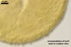
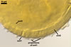
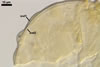
NOTES.
The distinctive feature of Gl. pustulatum
is the ornamentation of the outermost spore wall layer. The predominate structures
forming the ornamentation are blister-like outgrowths. In a cross view, they
are most frequently circular or arched. The size of these structures seems
to be inversely related to their frequency of occurrence on the spore surface.
Cup-shaped and irregular outgrowths occur rarely and mainly on spores densely
covered with blister-like thickenings. In a few specimens, both the number
of pustules and their size are very low and, hence, such spores are almost
smooth. The pustules are solid, not fatty, and do not disappear when spores
are mounted in lactic acid-based mountants.
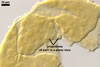
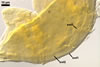
The
spore wall layer 2 easily separates into single laminae or their groups, especially
in vigorously crushed spores. The outer laminae are usually darker than the
inner ones, which sometimes are almost hyaline. The spore wall layer 3 is
most difficult to see, as it always tightly adheres to layer 2 and is hyaline.
Even vigorous crushing of spores does not separate it completely from the
laminae layer. However, numerous wrinkles on the inner surface of layer 2
and small areas of this layer separated from layer 2 supports its presence.
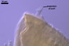
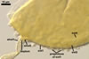
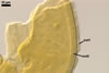
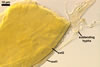
Most
spores of Gl. pustulatum recovered by the author of this website
possess a straight and cylindric or slightly flared subtending hypha. Its
wall is thin and usually lighter coloured than the spore wall. The pore in
the subtending hypha is usually closed by the thickened spore wall layer 2;
in a few spores, a curved septum continuous with the innermost lamina of spore
wall layer 2 is also present. According to Koske et al. (1986), the pore in
Gl. pustulatum is closed only by ingrowth of spore wall layer 2.
Alternative occlusion of a subtending hyphal pore either by wall thickening
or a septum has been described in other species of the genus Glomus
(Morton 1988).
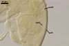
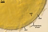
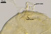
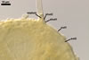
The spore
base of many specimens found is obscured by germ tubes growing most frequently
near the subtending hypha, although germinating spores with germ tubes developed
at the opposite of the subtending hypha are also present. The germ tubes grow
through a small hole seen in the spore wall layer 1. No spore germinating
by regrowth of the subtending hypha was found. This kind of germination most
frequently occurs in Glomus spp. (Morton and Benny 1990).
When
observed under a dissecting microscope, spores of Gl. pustulatum
resemble those of Gl. aggregatum
N.C. Schenck & G.S. Sm. emend. Koske, Gl.
arenarium Blaszk. et al., Gl.
claroideum N.C. Schenck & G.S. Sm., Gl. etunicatum W.N. Becker & Gerd., and Gl. hoi S.M. Berch & Trappe. Spores of all these species are yellow-coloured and have
a similar size (Berch and Trappe 1985; Blaszkowski et al. 2001; Schenck and
Smith 1982; Stürmer
and Morton 1997; Walker
and Vestberg 1998).
Examination
of the subcellular structure of the spores under a compound microscope readily
separates them. Only Gl. pustulatum produces spores whose the outermost
wall layer is permanent and ornamented with blister-like processes. In the
other fungi, the outermost spore wall layer sloughs and rarely occurs at maturity.
Glomus
trimurales Koske & Halvorson forms spores ornamented with blisters (Blaszkowski et
al. 2003). However, in contrast to those of Gl. pustulatum,
the blisters deteriorate and disappear with age.
REFERENCES
Berch S. M., Trappe J.
M. 1985. A new species of Endogonaceae, Glomus hoi. Mycologia 77, 654-657.
Blaszkowski J. 1994.
Polish Glomales 11. Glomus pustulatum. Mycorrhiza 4, 201-207.
Blaszkowski J. 1995.
Glomus corymbiforme, a new species in Glomales from Poland. Mycologia
87, 732-737.
Blaszkowski J., Adamska
I., Czerniawska B. 2003. Glomus trimurales, an arbuscular mycorrhizal
fungus (Glomerales) new for Poland and Europe. Mycotaxon 87, 425-436.
Blaszkowski J., Tadych
M., Madej T. 2001. Glomus arenarium, a new species in Glomales (Zygomycetes).
Acta Soc. Bot. Pol. 70, 97-101.
Blaszkowski J., Tadych
M., Madej T. 2002. Arbuscular mycorrhizal fungi (Glomales, Zygomycota) of
the Bledowska Desert, Poland. Acta Soc. Bot. Pol. 71, 71-85.
Dalpé Y. 1989.
Inventaire et repartition de la flore endomycorhizienne de dunes et de rivages
maritimes du Québec, du Nouveau-Brunswick et de la Nouvelle-Ecosse.
Naturaliste Can. (Rev. Ecol. Syst.) 116, 219-236.
Koske R. E., Friese C.,
Walker C., Dalpé Y. 1986. Glomus pustulatum: A new species
in the Endogonaceae. Mycotaxon 26, 143-149.
Mohankumar V., Ragupathy
S., Nirmala C. B., Mohadevan A. 1988. Distribution of vesicular-arbuscular
mycorrhizae (VAM) in the sandy beach soils of Madras coast. Cur. Sci. 57,
367-368.
Morton J. B. 1988. Taxonomy
of VA mycorrhizal fungi: classification, nomenclature, and identification.
Mycotaxon 32, 267-324.
Morton J. B., Benny G.
L. 1990. Revised classification of arbuscular mycorrhizal fungi (Zygomycetes):
a new order, Glomales, two new suborders, Glomineae and Gigasporineae, and
two new families, Acaulosporaceae and Gigasporaceae, with an emendation of
Glomaceae. Mycotaxon 37, 471-491.
Schenck N. C., Smith
G. S. 1982. Additional new and unreported species of mycorrhizal fungi (Endogonaceae)
from Florida. Mycologia 74, 77-92.
Stürmer S. L., Morton
J. B. 1997. Developmental patterns defining morphological characters in spores
of four species in Glomus. Mycologia 89, 72-81.
Tadych M., Blaszkowski
J. 2000a. Arbuscular fungi and mycorrhizae (Glomales) of the Slowinski National
Park, Poland. Mycotaxon 74, 463-483.
Tadych M., Blaszkowski
J. 2000b. Arbuscular mycorrhizal fungi of the Brda river valley in the Tuchola
Forests. Acta Mycol. 35, 3-23.
Walker C., Vestberg M.
1998. Synonymy amongst the arbuscular mycorrhizal fungi: Glomus claroideum,
G. maculosum, G. multisubstensum and G. fistulosum.
Ann. Bot. 82, 601-624.