Pacispora robiginia

Sieverd. & Oehl


In PVLG

SPORES produced singly in the soil, blastically at the tip of mycorrhizal extraradical hyphae. Spores pale orange (5A3) to golden yellow (5B7); globose to subglobose; (100-)125-155(-161) µm diam; rarely ellipsoidal; 95-135 x 135-165 µm; with a single subtending hypha.


SUBCELLULAR STRUCTURE OF SPORES consists of a spore wall and an inner germination wall.

In PVLG
In PVLG+Melzer's reagent

Spore wall consists of three layers (swl1-3).

Layer 1, forming the spore surface, permanent, of a smooth upper surface, unit, pale orange (5A3) to golden yellow (5B7), 2.5-3.5(-5.0) µm thick, tightly adherent to layer 2.

Layer 2 laminate, light orange (5A4-5), 4.0-6.5 µm thick.

Layer 3 permanent, concolorous with layer 2, <0.5 µm thick, usually tightly adherent to the lower surface of layer 2 in even vigorously crushed spores and, hence, very difficult to observe.

Germination wall includes three hyaline layers (gwl1-3).

Layer 1 flexible, 0.4-0.8 µm thick, usually separates from layer 2 in crushed spores.

Layer 2 coriaceous, 2.2-3.0 µm thick.

Layer 3 flexible, <1.0 µm thick, usually tightly adherent to the lower surface of layer 2, sometimes wrinkles in spores vigorously crushed in PVLG.

In Melzer's reagent, only layer 2 of the germination wall stains pare red (9A3).


GERMINATION SHIELD. Not found.


In PVLG
In PVLG+Melzer's

SUBTENDING HYPHA light orange (5A4-5) at the spore base and for some distance from the spore base; usually straight, sometimes slightly curved; cylindrical; 8-15 µm wide at the spore base, 12-25 µm at a some distance from the spore base.

Wall of subtending hypha light orange (5A4-5) for some distance from the spore base, then gradually lightens up to hyaline; 2.6-3.8 µm thick at the spore base, <1 µm thick 30-80(-100) µm below the spore base, composed of two layers continuous with spore wall layers 1 and 2.

Pore closed at the spore base by a transverse septum formed by spore wall layer 2 and by adherent spore wall layer 3.


GERMINATION. Unknown.


MYCORRHIZAE. Pacispora robiginia has been associated with mycorrhizal roots of plants of grasslands (Oehl and Sieverding 2004). However, attempts to grow this fungus in one-species cultures failed and, hence, the properties of mycorrhizae of P. robiginia remain unknown.


DISTRIBUTION. The type of P. robiginia has been selected from spores isolated from a calcareous Lithic Leptosol in the High Alpines at the Haldensteiner Calanda (9o27'E, 46o53'N) at 2800 m above the see level, near Chur (Kanton Graubünden), Switzerland (Oehl and Sieverding 2004). The same scientists have also found this fungus among roots of plants of High Alpine grasslands located in the Gotthard region (Kanton Wallis), Central Switzerland. The author of this website has recognized P. robiginia among spores extracted from under plants of the Tatra Mountains (Blaszkowski, unpubl. data).

NOTES. The morphological and biochemical properties of spores of P. robiginia presented above were prepared based on the original description of this fungus (Oehl and Sieverding 2004) and observations of its spores loaned from Dr. F. Oehl, Institute of Botany, University of Basel, Switzerland.

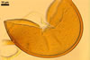
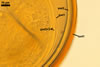
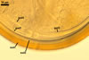
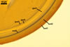
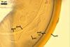
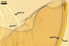
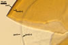
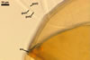
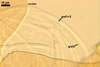
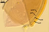
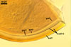
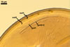
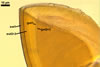
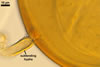
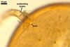
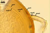
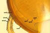
Under a dissecting microscope, spores of P. robiginia resemble yellow-coloured spores of many species of the genus Glomus. Examination of the subcellular structure of spores of the former fungus under a compound microscope readily reveals its main generic character, i. e., the inner complex and relatively thick germination wall resembling a germination wall of spores of fungi of the genus Scutellospora C. Walker & F.E. Sanders (Blaszkowski 2003). However, spores of Pacispora spp. form at the tip of more or less cylindrical hyphae, and thus identically to those of Glomus spp., whereas spores of the genus Scutellospora origin from a bulbous sporogenous cell.

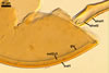
Of the known species of the genus Pacispora, only P. boliviana Sieverd. & Oehl forms spores of a similar colour to that of spores of P. robiginia (Oehl and Sieverding 2004). However, the upper surface of the structural laminate wall layer of spores of P. robiginia is smooth, and that of spores of P. boliviana is ornamented with shallow, usually pentagonal pits (Oehl and Sieverding 2004). Spores of the other species of this genus are colourless (Blaszkowski 2003; Oehl and Sieverding 2004).


REFERENCES

Blaszkowski J. 2003. Arbuscular mycorrhizal fungi (Glomeromycota), Endogone, and Complexipes species deposited in the Department of Plant Pathology, University of Agriculture in Szczecin, Poland. http://www.agro.ar.szczecin.pl/~jblaszkowski/.

Oehl F., Sieverding E. 2004. Pacispora, a new vesicular arbuscular mycorrhizal fungal genus in the Glomeromycetes. J. Appl. Bot. 78, 72-82.