DEVELOPMENT OF SPORES AND CHARACTERS OF MYCORRHIZAE
OF FUNGI OF THE GENUS PACISPORA
 |
 |
In PVLG |
|
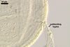
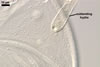
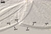
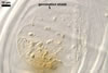
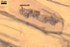
Spores of fungi of the genus Pacispora Sieverd. & Oehl. develop blastically at the end of cylindrical sporogenous hyphae (subtending hyphae) continuous with extraradical hyphae of arbuscular mycorrhizae. Thus, they develop identically as spores of fungi of the genus Glomus.
 |
 |
 |
 |
 |
 |
In PVLG |
In PVLG+Melzer's |
In PVLG
|
|||
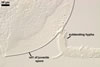
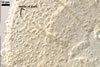
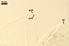
The wall of the most juvenile spores of members of this genus probably consists of only one layer (swl1), continuous with a one-layered wall of their subtending hypha, although the ontogenetic development of spores has been recognized only in P. scintillans (S.L. Rose & Trappe) Sieverd. & Oehl (Walker et al. 2004). The surface of this layer, forming the spore surface, is smooth (P. boliviana Sieverd. & Oehl, P. franciscana Sieverd. & Oehl, P. robiginia Sieverd. & Oehl) or ornamented with long [P. chimonobambusae (C.G. Wu & Y.S. Liu) Sieverd. & Oehl] or short [P. coralloidea Sieverd. & Oehl, P. scintillans] processes (Oehl and Sieverding 2004; Walker et al. 2004; Wu et al. 1995). Except for P. boliviana, this outermost spore wall layer is a permanent structure. In P. boliviana, this layer sloughs with age. After the full differentiation of the first layer, whose usually accompanies the termination of the spore expansion, a second laminate layer begins to form (swl2). When it is completely developed, a third layer origins (swl3). The third layer in all the species of Pacispora so far recognized is thin, concolorous with the laminate layer 2, to which it tightly adheres, and, thereby, it is very difficult to observe. Except for P. boliviana, the outer surface of the laminate layer of the other species of this genus is smooth (vs. pitted in P. boliviana).
 |
 |
 |
In PVLG
|
||
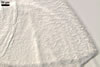
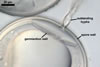
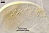
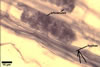
The next stage of the ontogenesis of spores of the genus Pacispora is the origination of a three-layered inner germination wall. The outermost layer 1 origins first (gwl1). It is thin, flexible, and hyaline. The middle layer 2 (gwl2) is hyaline, relatively thick, coriaceous sensu Walker (1986), and stains red to purple in Melzer's reagent. The innermost layer 3 (gwl3) is thin, hyaline, flexible, and tightly adheres to layer 2. The germination wall has no physical contact with the spore wall and origins independently and de novo after the full differentiation of the spore wall, similarly as in all members of the family Acaulosporaceae and the genus Scutellospora.
The ontogenetic development of spores of Pacispora spp. ends the formation of a uniform, plate-like germination shield on the surface of layer 1 of the inner germination wall. A germ tube grows from this shield and penetrates through the spore wall. After germination, the germination shield gradually deteriorates and then usually completely disappears. Hence, it is rarely visible in mature and older spores, similarly as in members of the family Acaulosporaceae.
 |
 |
 |
 |
 |
In PVLG
|
||||
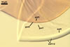
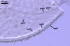
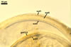
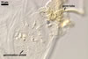
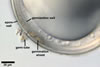
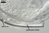
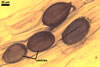
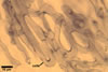
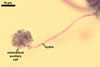
The mycorrhizae of P. scintillans, the only species of this genus known to form them, established from a single spore in roots of Plantago lanceolata L. consisted of arbuscules, vesicles, intra- and extraradical hyphae, as well as of auxiliary cells. The arbuscules, vesicles, and hyphae morphologically resembled those of Glomus spp. and stained intensively in 0.1% trypan blue. The auxiliary cells occurred both outside and inside roots, were knobby, and, thereby, similar to those formed by fungi of the genus Scutellospora.
 |
 |
 |
 |
 |
 |
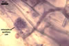 |
Arbuscules, vesicles, hyphae, extra- and intraradical auxiliary cells in and associated with roots of P. lanceolata
|
||||||
REFERENCES
Walker C. 1986. Taxonomic concepts in the Endogonaceae. II. A fifth morphological wall type in endogonaceous spores. Mycotaxon 25, 95-99.
Walker C., Blaszkowski J., Schwazott D., Schüßler A. 2004. Gerdemannia gen. nov., a genus separated from Glomus, and Gerdemanniaceae fam. nov., a new family in the Diversisporales based on the former Glomus scintillans. Mycol. Res. 108(6), 707-718.
Wu C.-G., Liu Y.-S., Hwuang Y.-L., Wang Y.-P., Chao C.-C. 1995. Glomales of Taiwan: V. Glomus chimonobambusae and Entrophospora kentinensis, spp. nov. Mycotaxon 53, 283-294.