DEFINITIONS OF MORPHOLOGICAL CHARACTERS OF SPORES
OF ARBUSCULAR FUNGI
The definitions of spore morphological and biochemical characters presented below are based
on the data proposed by Walker (1983, 1986), Morton (1986, 2002), Spain et al. (2006), and
those
received by the author of this website.
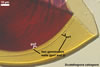
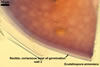
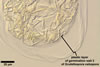
Spores of arbuscular fungi contain structures composed of component parts, of which each has unique phenotypic properties. According to Morton (2002), each spore, regardless of species, forms one spore wall. Additionally, all species of the genera Acaulospora, Ambispora, Archaeospora, Entrophospora, Gigaspora, Intraspora, Kuklospora, Pacispora, and Scutellospora produce spores with 1-3 inner walls called “germinal walls”. However, only in Gigaspora spp., the germinal wall is physically associated with their spore wall. In spores of the other genera, germinal walls origin and function independently on their spore wall.
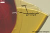
SPORE WALL. The spore wall originates from a sporogenous hypha. It develops at the tip of this hypha (Diversispora, Glomus, Gigaspora, Pacispora, Paraglomus, and Scutellospora spp.), inside it (Entrophospora, Intraspora, Kuklospora, and some Glomus spp. producing spores intercalary), or from its side (Acaulospora, Ambispora, Archaeospora, and Otospora spp.). This layer grows, thickens, and differentiates its components along with the increasing diameter of a spore. The differentiation of the spore wall components stops when the spore has ceased to expand. Later, some small changes in colour, thickness, and rigidity of this wall may be observed.
The spore wall of the species of arbuscular fungi recognized consists of at least two layers, although some species differentiate even a 4-layered spore wall. The wall of the most juvenile spores may be 1- or 2-layered. Except for Ap. gerdemannii, the spore wall layers originate successively towards the spore interior. In Ap. gerdemannii, spore wall layer 2 forms first and then an outer layer origins de novo (Morton and Redecker 2001).
The layers of a spore wall and germinal walls may differ in both phenotypic and biochemical properties.
Eight phenotypes of spore wall layers were recognized to date.
 |
 |
 |
In PVLG+Melzer's reagent |
||
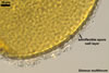
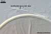
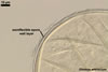
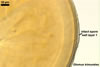
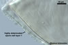
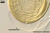
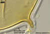
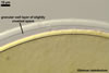
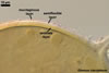
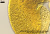
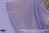
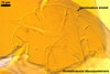
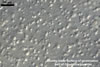
1. Mucilagenous layer. This layer forms the spore surface of some Glomus spp., e. g. Gl. claroideum, Gl. mosseae, and Gl. multiforum. In juvenile spores, this layer is smooth or somewhat roughened. When present, it stains intensively red in Melzer’s reagent. However, it quickly deteriorates and sloughs, even in young spores, and, thereby, rarely occurs in the spore wall structure of mature spores.
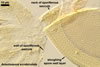
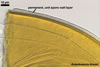
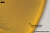
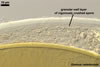
2. Semiflexible layer. A semiflexible layer usually is thin, but somewhat less flexible than thin, flexible layers present in inner germinal walls of species of the genera, e. g., Acaulospora and Scutellospora. In crushed specimens, the semiflexible layer frequently detaches from a layer adherent to its lower surface and does not wrinkle as a flexible layer (see, e. g., Gl. arenarium). When the semiflexible layer covers a laminate spore wall layer, its longevity is higher than that of a mucilaginous layer, although this layer rarely is present intact or at all in mature spores. Generally, in species having a semiflexible layer, it is smooth when not deteriorated. In Gl. multiforum, the lower surface of the semiflexible layer is ornamented with ingrowths being the base to form pits in the upper surface of the next laminate layer. In all species recognized to have a semiflexible layer in the structure of their spores, this layer does not react in Melzer’s reagent.
 |
 |
 |
 |
In PVLG |
In PVLG+Melzer's |
||
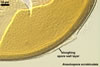
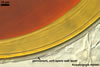
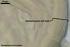
3. Other deteriorating and sloughing layers. These layers form a spore surface and are more or less persistent. In young and sometimes in mature spores, these layers resemble a unit layer. However, with time, these layers always deteriorate and frequently are absent in older spores. In some species, e. g., in Gl. trimurales, such a layer is ornamented with blister-like processes that deteriorate and disappear with time. In Acaulospora, Appendicispora, Archaeospora, Entrophospora, Intraspora, Kuklospora, and Otospora spp., the sloughing layer is continuous with the wall of the neck of a sporiferous saccule.
 |
 |
 |
 |
 |
 |
 |
In PVLG+Melzer's |
In PVLG |
In PVLG+Melzer's
reagent |
In PVLG |
|||
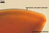
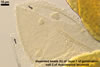
Another layer deteriorating and sloughing with age is a layer expanding in lactic acid-based mountants. It was originally found in the wall structure of spores of Gl. pansihalos (Berch and Koske 1986). This phenotype also occurs in spores of A. morrowiae and Gl. arenarium (Blaszkowski et al. 2001; Morton 2000).
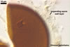
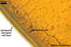
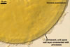
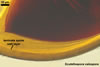
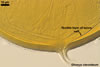
4. Unit layer. This layer is a uniform and persistent structure. It may be colourless, as, e. g., in A. thomii, or coloured, as, e. g., in G. gigantea. In some species, this layer is ornamented with different processes (e. g., with warts, as in S. persica). Sometimes, the unit layer is very thin and similarly coloured as its penultimate layer, as, e. g., in S. pellucida. Hence, this layer may be easily omitted. In Melzer’s reagent, the unit layer usually is nonreactive or its staining reaction is less intensive than that of other layers. In the species recognized to date, this layer forms a spore surface.
 |
 |
 |
 |
 |
 |
 |
In PVLG |
In PVLG+Melzer's |
In PVLG |
In PVLG+Melzer's |
|||
 |
 |
In PVLG |
|
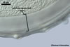
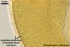
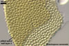
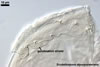
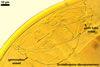
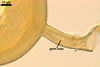
6. Laminate layer. A laminate layer is a conglomerate of usually very thin, <0.5 µm, tightly adherent sublayers. In vigorously crushed spores, groups of sublayers or the innermost sublayer sometimes separate(s) from the conglomerate. This may lead to an erroneous conclusion of the presence of additional layers. In a few species, e. g., in Paraglomus laccatum, the sublayers are thick and easily separate from each other. In the most juvenile spores, a laminate wall represents a single sublayer. With time, next sublayers are added until the spore ceases to expand. The laminate layer may be colourless through the entire life of a fungus, as, e. g., in Par. laccatum and Gl. diaphanum, or may gradually darken because of the addition of next, coloured sublayers. Both the upper and the lower surface of the laminate layer may be smooth or ornamented with processes (e. g., E. infrequens and Gl. pansihalos) or pits (e. g., A. cavernata, A. lacunosa, Gl. insculptum, and Gl. multiforum). In species of the genera Diversispora, Gigaspora, Glomus, Pacispora, Paraglomus, and Scutellospora, the laminate layer is continuous with the laminate layer of the subtending hypha or the sporogenous cell. Except for members of the genera Acaulospora and Entrophospora, the spore wall of species of the other genera of the Glomeromycota contains only one laminate layer. In most Acaulospora spp., their 3-layered spore wall contains two inner laminate layers. In the spore wall of E. infrequens, its second layer also is finely laminate. The permanent laminate layer generally is the main structure protecting the spore inside from the influence of different abio- and biotic stresses. In E. infrequens, the second laminate spore wall layer deteriorates with age and is frequently completely sloughed in mature spores.
 |
 |
 |
 |
 |
 |
 |
In PVLG+Melzer's reagent |
In
PVLG |
In PVLG+Melzer's |
In PVLG |
|||
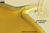
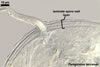
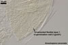
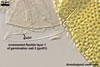
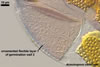
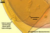
7. Flexible layer. This phenotype is a thin, ca. 0.5 µm thick, and flexible structure, usually wrinkling in spores crushed in PVLG. The layer resembles flexible layers of germinal walls of spores of fungi of the genera Acaulospora, Ambispora, Archaeospora, Entrophospora, Intraspora, Kuklospora, Otospora, Pacispora, and Scutellospora.
 |
 |
 |
 |
In PVLG+Melzer's |
In PVLG
|
In PVLG+Melzer's |
|
However, it originates from the inner surface of the subtending hyphal wall continuous with the spore wall, and not independently on the spore wall, as in in spores of the genera listed above. The flexible layer occurs in the wall structure of spores of, e. g., Gl. claroideum and Gl. lamellosum. In the latter species, the flexible layer stains pinkish white to pastel red in Melzer’s reagent.
 |
 |
In PVLG |
In PVLG+Melzer's |
 |
 |
 |
 |
In PVLG+Melzer's
|
In PVLG |
||
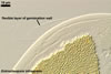
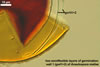
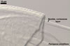
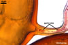
GERMINAL WALL. Germinal walls produce all fungi of the genera Acaulospora, Ambispora, Archaeospora, Entrophospora, Gigaspora, Intraspora, Kuklospora, Otospora, Pacispora, and Scutellospora. The number of germinal walls in the species described ranges from 1 to 3. Each wall consists of 1-3 layers. The first germinal wall forms after the synthesis of the spore wall has finished and it has no physical connection with this wall. The next wall originates similarly, but only after the first one has completed differentiation of all its layers. The germinal walls of all species are colourless. In most taxa forming germinal walls, their components are more or less flexible. The full differentiation of germinal wall(s) condition(s) the acquirement of the ability of a spore to germinate. In members of the genus Gigaspora, a germ tube directly originates from the germinal wall tightly adherent to their spore wall. In contrast, in species of the other genera forming at least one germinal wall, germ tubes grow from earlier produced pre-germination structures. They are called "germination orb" (Acaulospora and Kuklospora spp.), "germination shield" (Pacispora and Scutellospora spp.), or "germination structure" (Ap. appendicula). These pre-germination structures are always placed on the upper surface of the innermost germinal wall. No pre-germination structures have been detected in spores of Entrophospora, Intraspora, and Otospora spp. to date.
In the germinal walls of the species of arbuscular fungi described, six phenotypes of layers were revealed.
 |
 |
 |
In PVLG |
In PVLG+Melzer's |
|
1. Smooth, thin, flexible layer. This layer is smooth, thin, ca. 0.5 µm thick, and wrinkles in spores crushed in PVLG. In most arbuscular fungi, this layer co-occurs with a tightly adherent second layer of identical properties. Hence, such a 2-layered structure may be erroneously interpreted as one layer. Two adherent layers of the same thickness usually occur in the first germinal wall, when at least one next germinal wall is produced. In fungi forming only one germinal wall, this wall may consist of two, e. g., S. persica, or three, e. g., P. scintillans, layers, of which the outermost one always is thinner than the middle or the innermost layer. The smooth, flexible, membranous layer sometimes stains pale in Melzer’s reagent.
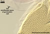
2. Ornamented, thin, flexible layers. Two phenotypes of ornamented, thin, flexible, layers were so far revealed: a beaded layer and a knobby layer. The beaded layer commonly occurs in the structure of the innermost germinal wall of spores of the genera Acaulospora and Kuklospora. It is thin, ca. 0.5 µm thick, and covered with granular excrescences (beads) that frequently disperse in crushed spores. The beaded layer always covers a highly flexible (plastic) layer staining intensively red to purple in Melzer’s reagent.
 |
 |
 |
In PVLG |
||
The knobby layer is the outermost one in the 3-layered innermost germinal wall of spores of A. cavernata. This layer is evenly ornamented with small knobs. The knobs do not disperse in crushed spores. The knobby layer resembles a plastic covering of the lower surface of post envelopes. It is associated with a flexible layer, which does not stain in Melzer’s reagent.
A similar knobby layer represents the 1-layered germinal wall of G. gigantea spores. However, the knobs of this layer grow inward the spore (vs. they ornament the upper surface of layer 1 of the germinal wall 2 of A. cavernata spores).
 |
 |
 |
 |
In PVLG
|
In PVLG+Melzer's
|
||
3. Thin, semiflexible layer. In contrast to a flexible layer, the semiflexible layer is somewhat more rigid and does not wrinkle in crushed spores. However, this layer does not crack as a thin, rigid layer characterized below. The semiflexible layer occurs alone or adheres to another semiflexible layer in some species of the genera Acaulospora, e. g. in A. mellea and A. scrobiculata, and Kuklospora.
4. Thick, flexible layer. Walker (1986) called it “coriaceous wall”. It was described as having a leather-like appearance in hypertonic solutions. This layer occurs, e. g., in the middle of the 3-layered innermost germinal wall of spores of A. cavernata and P. scintillans and usually is the penultimate layer in the innermost germinal wall of many species of the genus Scutellospora. This layer sometimes stains pale in Melzer’s reagent.
5. Plastic layer. Morton (1986) described a plastic layer as “an amorphous wall”. This layer is highly plastic with applied pressure to spores crushed in lactic acid-based mountants. The plastic layer always is the innermost layer in the subcellular structure of spores and stains dark red to red-purple in Melzer’s reagent. It is present in many species of the genera Acaulospora, Kuklospora, and Scutellospora.
 |
 |
In PVLG+Melzer's
|
|
 |
 |
 |
In PVLG+Melzer's reagent |
In PVLG |
|
6. Thin, rigid layer. A thin, rigid layer forms the first, 1-layered germinal wall of spores of A. gedanensis. Such layers also occur in the middle, 2-layered germinal wall of spores of Ap. appendicula, Ap. fennica, and Ap. gerdemannii. In Ac. gedanensis, Ap. fennica, and Ap. gerdemannii, these layers are smooth, and in Ap. appendicula they are ornamented with a convex, alveolate reticulum. Unlike all the other species of arbuscular fungi producing spores with flexible or semiflexible germinal walls, this type of layer is fragile in all the species listed here.
PRE-GERMINATION STRUCTURES. Three types of pre-germination structures, from which germ tubes arise, were recognized to date.
 |
 |
In PVLG |
|
 |
 |
In PVLG |
|
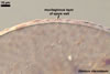
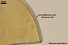
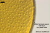
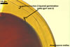
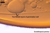
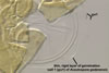
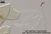
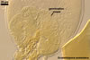
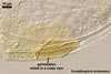
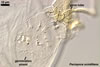
1. Germination orb. A germination orb is formed by a centrifugally rolled, hyaline to light-coloured hypha. The orb usually is circular or somewhat elliptic when seen in a plane view and located on the upper surface of the innermost germinal wall when observed in a cross view. The germination orb is an impermanent structure. With time, it decomposes and may be completely unrecognizable in older spores. Germination orbs were found only in a few species of the genus Acaulospora and Kuklospora.
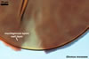
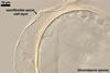
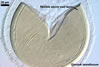
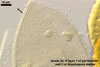
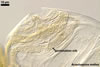
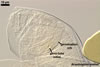
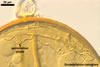
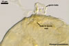
2. Germination shield. Except for the germination shield of spores of S. projecturata, the germination shields of the other species of the genus Scutellospora are thin, uniform, elliptic or cardioid, hyaline to coloured lobes with a more or less incurved or incised margin. They commonly occur in mature spores of Scutellospora spp. The formation of this structure on the upper surface of the innermost germinal wall ends the ontogenetical development of spores of these fungi. The germination shields do not deteriorate with age. In S. projecturata, the germination shield is formed by a coiled hypha and resembles germination orbs of Acaulospora and Kuklospora spp. (Kramadibrata et al. 2000).
 |
 |
In PVLG |
|
 |
 |
 |
 |
 |
 |
 |
In PVLG
|
In PVLG+Melzer's reagent
|
|||||
 |
 |
 |
 |
In PVLG |
|||
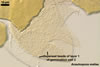
the other members of the phylum Glomeromycota, only P. scintillans and P. franciscana were found to form germination shields resembling in appearance those produced by Scutellospora spp. However, the germination shields in these species decompose with age and usually are invisible in older spores.
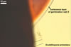
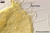
3. Germinal wall. A germinal wall is a semiflexible layer from which a germ tube emerges. This layer is ornamented with knobs distributed on its lower surface. Once it is differentiated, it remains unchanged as long as a spore retains.
|
 |
 |
In PVLG |
||
4. Germination structure. Such a structure, resembling a germination shield of spores of fungi of the genus Scutellospora, has been detected on the upper surface of the second germinal wall of spores of Ap. appendicula (Spain et al. 2006). Unfortunately, there is no mention of its persistency.
When germination was observed, a germ tube of Glomus spp. grew from either the inner surface of the subtending hyphal wall near the spore, a septum of the subtending hypha, or from the spore wall. In all the cases, no distinct pre-germination structure was revealed.
 |
 |
 |
 |
In PVLG |
In PVLG+Melzer's |
In PVLG |
|
REFERENCES
Berch S. M., Koske R. E. 1986. Glomus pansihalos: a new species in the Endogonaceae, Zygomycetes. Mycologia 78, 838-842.
Blaszkowski J., Tadych M., Madej T. 2001. Glomus arenarium, a new species in Glomales (Zygomycetes). Acta Soc. Bot. Pol. 70, 97-101.
Kramadibrata K., Walker C., Schwarzott D., Schüßler A. 2000. A new species of Scutellospora with a coiled germination shield. Ann. Bot. 86, 21-27.
Morton J. B. 1986. Three new species of Acaulospora (Endogonaceae) from high aluminum, low pH soils in West Virginia. Mycologia 78, 641-648.
Morton J. B. 2002. International Culture Collection of Arbuscular and Vesicular-Arbuscular Mycorrhizal Fungi. West Virginia University.
Morton J. B., Redecker D. 2001. Two families of Glomales, Archaeosporaceae and Paraglomaceae, with two new genera Archaeospora and Paraglomus, based on concordant molecular and morphological characters. Mycologia 93, 181-195.
Oehl F., Sieverding E. 2004. Pacispora, a new vesicular arbuscular mycorrhizal fungal genus in the Glomeromycetes. J. Appl. Bot. 78, 72-82.
Spain J. L., Sieverding E., Oehl F. 2006. Appendicispora: a new genus in the arbuscular mycorrhiza-forming Glomeromycetes, with a discussion of the genus Archaeospora. Mycotaxon 97, 163-182.
Walker C. 1983. Taxonomic concepts in the Endogonaceae: spore wall characteristics in species descriptions. Mycotaxon 18, 443-455.
Walker C. 1986. Taxonomic concepts in the Endogonaceae. II. A fifth morphological wall type in endogonaceous spores. Mycotaxon 25, 95-99.